
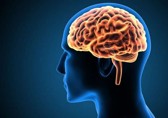
The brain is an astoundingly complex somatic machine. It generates electricity, records data, processes information, and operates an intricate system of nerves that control physical movement. Over the years medical professionals and scientists have unlocked many secrets about the human brain, but for every discovery, there have been even more questions. Modern technology has attempted to answer these questions by creating new ways to study and map the brain. These techniques help scientists better understand the complexities of the mind and assist doctors in finding ways to treat it.
If you are a student of the mind, or a patient seeking treatment, you have probably heard of brain scanning. The enterprise is at the forefront of Neurology. Using various techniques the brain is mapped and its processes recorded. From the information gathered neurologists can better understand the analytical workings of your mind. Mapping allows medical professionals to better understand mental conditions and creates new ways for them to be treated. Here are the three main types of brain imaging used by the medical profession today.
Electroencephalography (EEG)
In an EEG, electrodes are placed about the face and scalp. The electrodes take measurements so they have to be placed in correct positions in order to be effective. Once in place, the electrodes will study a person’s mental state. The patient in question follows a certain action like sleeping, walking, or even waking from sleep. The EEG maps the signals coming from the electrodes and then technicians study the data.
EEG is great at studying activity in certain parts of the brain and has been proven effective for the treatment of seizures. Its only drawback is that electrical measurements differ so greatly from person to person makes it difficult to decipher the region signals are coming from.
Positron Emission Tomography (PET)
PET scans measure sugar glucose levels in the brain. This allows them to discover the location of neural firing. A PET scan begins with the injection of a tracer substance into the blood. The tracer has radioactive isotopes attached to it. As certain parts of the brain activate the blood containing the tracer transports oxygen. The tracer leaves spots visible by detectors that create a video image. This allows technicians to observe brain activity during specific tasks.
PET scans are limited to generalized areas of the mind. This presents a drawback as they cannot be overly specific. However, despite their invasive and costly manner, PET scans have been proved to use in the medical diagnosis of serious conditions. Chief among these conditions Alzheimer’s.
Magnetic Resonance Imaging (MRI)
An MRI is the most popular form of brain scanning. Many people are familiar with the name, the process, and have seen a slew of them performed in movies. This is mainly because brain MRI information is very useful for psychology.
An MRI utilizes strong magnetic fields that align spinning atomic nuclei found in body tissues. The fields disturb these nuclei’s axis of rotation requiring them to return to their baseline status. This journey produces radio frequency signals that are then observed by technicians.
MRI scans posit little risk to health and are noninvasive. This allows them to be used for all manner of the individual despite age. MRIs can even be used on infants both born and still in utero. They provide constant imaging across the spectrum of development. An MRI is not a fun process as patients have to sit still in a claustrophobic tube for an extended period of time.
An MRI is useful for mapping signal frequencies within the brain. They can be used to detect a large variety of abnormalities in the brain including tumours. An MRI also provides a measurement of both function and structure.